
Antioxidant Effect of Hibiscus sabdariffa calyx Aqueous Extract on Mice Brains Exposed to MonosodiumGlutamate- Induced Alterations Effet antioxydant de l'extrait aqueux de calices d'Hibiscus sabdariffa sur les cerveaux de souris exposées à desaltérations induites par le glutamate monosodique
Article Sidebar
Abstract Views | PDF/EPUB Downloads
0
/ 0
/ 0
Main Article Content
Abstract
Background: Monosodium glutamate (MSG), a common flavor enhancer, has been linked to neurotoxicity and oxidative stress in the brain, contributing to neurodegenerative changes. Hibiscus sabdariffa calyx aqueous extract HSAE), rich in polyphenols and anthocyanins, possesses antioxidant properties that may counteract such effects. Objective: To evaluate the antioxidant and neuroprotective potential of HSAE against MSG-induced brain alterationsin mice.
Method: Male mice were divided into six groups (n=6): control, MSG (2.5 g/kg), fluoxetine (20 mg/kg), and HSAE (50, 100, and 200 mg/kg) co- administered with MSG for 14 days respectively. Oxidative stress markers including malondialdehyde (MDA), nitrites, glutathione (GSH), catalase (CAT), superoxide dismutase (SOD), and acetylcholinesterase were assessed, alongside histological examination of the dentate gyrus.
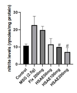
Result: MSG significantly increased MDA, nitrites, and acetylcholinesterase levels while decreasing GSH, CAT, and SOD, indicating oxidative damage and cholinergic dysfunction. HSAE (100 and 200 mg/kg) and fluoxetine significantly reversed these effects. Notably, HSAE (200 mg/kg) restoredantioxidant enzyme levels and neuronal integrity comparable to fluoxetine.
Conclusion: HSAE significantly demonstrated strong antioxidant and neuroprotective effects against MSG-induced neurotoxicity, particularly at 200 mg/kg, by reducing oxidative stress and preserving brain structure.
Résumé
Contexte : Le glutamate monosodique (GMS), un exhausteur de goût couramment utilisé, a été associé à une neurotoxicité et à un stress oxydatif cérébral, contribuant ainsi à des altérations neurodégénératives. L'extrait aqueux de calice d'Hibiscus sabdariffa (EACHS), riche en polyphénols et en anthocyanes, possède des propriétés antioxydantes susceptibles de contrecarrer ces effets.
Objectif : Évaluer le potentiel antioxydant et neuroprotecteur de l'EACHS contre les altérations cérébrales induites par le GMS chez la souris.
Méthode : Des souris mâles ont été réparties en six groupes (n=6) : un groupe témoin, un groupe GMS (2,5 g/kg), un groupe fluoxétine (20 mg/kg) et un roupe EACHS (50, 100 et 200 mg/kg) co-administré avec du GMS, pendant 14 jours. Les marqueurs du stress oxydatif, notamment le malondialdéhyde (MDA), es nitrites, le glutathion (GSH), la catalase (CAT), la superoxyde dismutase (SOD) et l'acétylcholinestérase, ont été évalués, parallèlement à un
examen histologique du gyrus denté.
Résultats : Le glutamate monosodique (GMS) a entraîné une augmentation significative des taux de MDA, de nitrites et d'acétylcholinestérase, tout en diminuant ceux de GSH, de CAT et de SOD, ce qui indique des lésions oxydatives et un dysfonctionnement cholinergique. L'extrait aqueux EACHS (100 et 00 mg/kg) et la fluoxétine ont significativement inversé ces effets. Notamment, l'extrait EACHS (200 mg/kg) a rétabli les taux d'enzymes antioxydantes et l'intégrité neuronale de manière comparable à la fluoxétine.
Conclusion : L'EACHS a démontré de manière significative de puissants effets antioxydants et neuroprotecteurs contre la neurotoxicité induite par le GMS, en particulier à la dose de 200 mg/kg, en réduisant le stress oxydatif et en préservant la structure cérébrale.
Downloads
Article Details
Issue
Section

This work is licensed under a Creative Commons Attribution-NonCommercial-NoDerivatives 4.0 International License.
How to Cite
Share
References
1.Davis CC, Choisy P (2024). Medicinal plants meet modern biodiversity science. Current Biology 34: R158-R173.
2.Ingale S, Rathored J, Wankhade S, Nibude S. (2025). Exploring the use of herbal medicines in the context of dermatokinetic research. Journal of Young Pharmacists 17: 19-25.
3.Takada K, Nakano S, Nishio R, Muku D, Mochizuki S, Inui I, Okita K, Koga A, Watanabe K, Yoshioka Y, Ariyoshi W, Yamasaki R (2024). Medicinalherbs, especially Hibiscus sabdariffa, inhibit oral pathogenic bacteria. Journal of Oral Biosciences 66:179-187.
4.Aliya F, Yunus MH, Izhar SK, Afaq U (2025). Medicinal plants in the treatment of respiratory diseases and their future aspects. Recent Patents on Biotechnology 19: 2-18.
5.Nuro G, Tolossa K, Arage M, Giday M (2024). Medicinal plants diversity among the Oromo community in Heban-Arsi district of Ethiopia used to manage human and livestock ailments. Frontiers in Pharmacology 15: 1455126.
6.Wachtel-Galor S, Benzie IFF (2011). Herbal medicine:An introduction to its history, usage, regulation, current trends, and research needs. In I. F. F. Benzie & S. Wachtel-Galor (Eds.), Herbal medicine: Biomolecular and clinical aspects; CRC Press.
7.Espinosa-Sánchez A, Montaño-Estrada LF, Rendón-Huerta EP, Navarrete-Peón A, Gómez-Aldapa CA, Muñóz-Pérez VM, Castro-Rosas J. (2024), Hibiscus sabdariffa as a Novel Alternative Strategy Against Helicobacter pylori Infection Development to Gastric Cancer. Journal of Medicinal Food 27(12):1158-1167.
8. Wang J, Cao X, Jiang H, Qi Y, Chin KL, Yue Y (2014). Antioxidant activity of leaf extracts from different Hibiscus sabdariffa accessions and simultaneous determination of five major antioxidant compounds by LC-Q-TOF-MS. Molecules, 19, 21226-21238.
9. Bernardes AL, Moraes LFS, Cruz BCDS, da Conceição LL, de Oliveira LL, Sarandy MM, Gonçalves RV, Peluzio MDCG (2023). Hibiscus sabdariffa supplementation increases butyrate synthesis and reduces inflammatory cells, attenuating theformation of aberrant crypt foci in BALB/c mice induced to pre-neoplastic lesions. British Journal of Nutrition 129: 352-363.
10. Rangel-García CA, Reynoso-Camacho R, Pérez- Ramírez IF, Morales-Luna E, de Los Ríos EA, Salgado LM (2022). Serum phospholipids are potential therapeutic targets of aqueous extracts of roselle (Hibiscus sabdariffa) against obesity and insulin resistance. International Journal of Environmental Research and Public Health 19: 16538.
11. Xu DP, Li Y, Meng X, Zhou T, Zhou Y, Zheng J, Zhang JJ, Li HB (2017). Natural antioxidants in foods and medicinal plants: Extraction, assessment and resources. International Journal of Molecular Sciences, 18: 96.
12. Ngondo B, Mpika J, Mbon N (2023). Antioxidant activity of phenolic compounds in Hibiscus sabdariffa from Congo. African Journal of Agricultural Research. 19: 1056-1068.
13. Kayode OT, Rotimi DE, Kayode AAA, Olaolu TD, Adeyemi OS (2020). Monosodium glutamate (MSG)- induced male reproductive dysfunction: A mini review. Toxics 8: 7.
14. Gbadamosi I, Yawson EO, Akesinro J, Adeleke O, Tokunbo O, Bamisi O, Ibrahim-Abdulkareem R, Awoniran P, Gbadamosi R, Lambe E, Atoyebi A, Bayo- Olugbami AA, Abayomi TA, Arogundade TT (2024). Vitamin D attenuates monosodium glutamate- induced behavioural anomalies, metabolic dysregulation, cholinergic impairment, oxidative stress, and astrogliosis in rats. Neurotoxicology, 103: 297-309.
15. Das D, Banerjee A, Manna K, Sarkar D, Shil A, Sikdar N, Bhakta M, Mukherjee S, Maji BK. (2023). Quercetin counteracts monosodium glutamate to mitigate immunosuppression in the thymus and spleen via redox-guided cellular signaling. Phytomedicine 126: 155226.
16. Nuhu F, Gordon A, Sturmey R, Seymour AM, Bhandari S (2020). Measurement of glutathione as a tool for oxidative stress studies by high performance liquid chromatography. Molecules 25: 4196. https://doi.org/10.3390/molecules25184196.
17. Moron MS, DePierre JW, Mannervik B (1979). Levels of glutathione, glutathione reductase and glutathione S-transferase activities in rat lung and liver. Biochimica et Biophysica Acta 582: 67-78.
18. Adeoluwa OA, Agboola I, Akinluyi E, Eduviere A, Adeoluwa AG, Ajayi A, Aderibigbe A (2023). N-butanol fraction of Olax subscorpioidea attenuates lipopolysaccharide-induced depressive-like symptoms in mice. Research Square, 7: 19.
19. Adam-Vizi V, Seregi M (1982). Receptor independent stimulatory effect of noradrenaline on Na+ /K+ - ATPase in rat brain homogenate: Role of lipid peroxidation. Journal of Neurochemistry 38: 1148- 1152.
20. Misra HP, Fridovich I (1972). The role of superoxide anion in the autoxidation of epinephrine and a simple assay for superoxide dismutase. Journal of Biological Chemistry 247: 3170-3175.
21. Green LC, Wagner DA, Glogowski J, Skipper PL, Wishnok JS, Tannenbaum SR (1982). Analysis of nitrate, nitrite, and [15N] nitrate in biological fluids. Analytical Biochemistry, 126: 131-138.
22. Góth L (1991). A simple method for determination of serum catalase activity and revision of reference range. Clinica Chimica Acta 196: 143-151.
23. Ellman GL, Courtney KD, Andres V Jr, Featherstone RM (1961). A new and rapid colorimetric determination of acetylcholinesterase activity. Biochemical Pharmacology 7: 88-95.
24. Eweka AO, Om'Iniabohs FAE (2007). Histological studies of the effects of monosodium glutamate on the small intestine of adult Wistar rats. Internet Journal of Gastroenterology 6(2).
25. Farombi EO, Onyema OO (2006). Monosodium glutamate-induced oxidative damage and genotoxicity in the rat: Modulatory role of vitamin C, vitamin E and quercetin. Human & Experimental Toxicology 25: 251-259.
26. Omogbiya AI, Ben-Azu B, Eduviere AT, Eneni AO, Nwokoye PO, Ajayi AM, Umukoro S (2020). Monosodium glutamate induces memory and hepatic dysfunctions in mice: Ameliorative role of Jobelyn®. Toxicology Research 37: 323-335.
27. Kong JM, Chia LS, Goh NK, Chia TF, Brouillard R (2003). Analysis and biological activities of anthocyanins. Phytochemistry 64: 923-933.
28. Da-Costa-Rocha I, Bonnlaender B, Sievers H, Pischel I, Heinrich M (2014). Hibiscus sabdariffa L.-A phytochemical and pharmacological review. Food Chemistry 165: 424-433.
29. Gad SB, Zaghloul DM, Abdel-Rahman GH (2020). Protective effect of Hibiscus sabdariffa extract against monosodium glutamate-induced testicular toxicity in rats: Role of oxidative stress and apoptosis. Journal of Complementary and Integrative Medicine 17: 20190135.
30. Farag MF, Osman N (2009). Antioxidant Actions of Irradiated Hibiscus sabdariffa L. (KARKADE) Against Monosodium Glutamate-Induced Oxidative Stress in Rats. Isotope and Radiation Research, 41.
31. Ezejiofor AN, Orisakwe OE, Obi E (2020). Antioxidant and hepatoprotective effects of Hibiscus sabdariffa aqueous extract against MSG-induced liver damage. Journal of Applied Pharmaceutical Science 10: 120-127.
32. Mahmoud RH, Mohamad HF (2014). Hibiscus sabdariffa L. attenuates nephrotoxicity and oxidative stress. American Journal of Food Technology 9(6): 299-307.
33. Ngulde SS, Usman H, Yahaya Y (2021). Antioxidant and neuroprotective effect of Hibiscus sabdariffa against MSG-induced brain damage in rats. Heliyon, 7(1).
34. Choi DW (2020). Glutamate neurotoxicity and diseases of the nervous system. Neuron, 106: 866-887.
35. Arias-Sánchez S, Vázquez-Medina JP, Fernández-García E (2023). Phytochemical composition and biological activities of Hibiscus sabdariffa L.: A review of its antioxidant and anti-inflammatory properties. Food Chemistry 405: 134824.
36. Duman RS, Aghajanian GK, Sanacora G, Krystal JH (2016). Synaptic plasticity and depression: new insights from stress and rapid-acting antidepressants. Nature Medicine 22: 238-249.
37. Kitahara Y, Tsurugizawa T, Uematsu A, Ogawa M, Kato F, Saito Y (2016). Chronic fluoxetine treatment increases hippocampal neurogenesis and brain-derived neurotrophic factor levels in adult mice. Neuroscience Research 110: 56-63.